Achilles calcific tendinitis
Achilles insertional calcific tendinopathy (ACIT)
Achilles Insertional Calcific Tendinopathy (ACIT) is a condition caused by the deterioration of the Achilles tendon in the heel, resulting in bone spurs. ACIT can cause heel pain in both active and inactive people and can be aggravated by activity or footwear. It does not happen instantly; it usually takes many years for ACIT to occur. Many treatment options are available for a quick and swift return to daily life. The condition is also called Insertional Achilles Tendinitis.
Anatomy

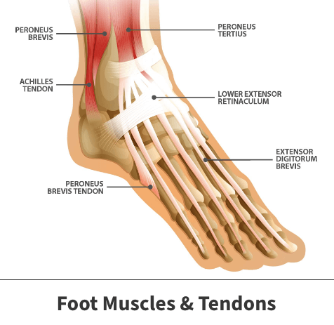
The Achilles tendon, also known as the calcaneal tendon, is a tough, fibrous band of tissue that connects the calf muscle to the heel bone (calcaneus). When the calf muscles flex, the Achilles tendon pulls on the heel, allowing you to walk, run, climb stairs, jump, and stand on the tip of your toes. It is both the strongest and largest tendon in the entire body. While it may be the strongest, it is also vulnerable to injury, due to the high tensions placed on it and its limited blood supply.
About
The Achilles tendon continually undergoes stress. When the tendon is not given enough time to recover, microscopic tearing begins to occur. Over time, the damaged tendon fibers start to harden (calcify), often times resulting in the formation of bone spurs. This deterioration and calcification is known as ACIT. ACIT can happen to both active and inactive patients and is not related to a specific injury. It comes from years of overuse or abuse. For example, long distance runners and sprinters are both known to suffer from ACIT. It can cause heal pain and can be aggravated by activity or footwear.
Additional factors resulting in ACIT include:
- A sudden increase in the amount and/or intensity of exercise activity—for example, increasing the distance you run every day by a few miles without giving your body a chance to adjust to the new distance
- Tight calf muscles—Having tight calf muscles and suddenly starting an aggressive exercise program can put extra stress on the Achilles tendon

Symptoms
Common symptoms of ACIT include:
- Stiffness and pain along the Achilles tendon that worsens with activity and is present in the morning
- Severe pain the day after exercising
- Thickening of the tendon
- Bone spurs
- Constant swelling that gets worse throughout the day with activity
- A sudden “pop” in the back of your calf or heel
Diagnosis
Your Florida Orthopaedic Institute physician will examine your foot and ankle, looking for the following signs:
- Swelling along the Achilles tendon or at the back of your heel
- Thickening or enlargement of the Achilles tendon
- Bony spurs at the lower part of the tendon at the back of your heel
- The point of greatest tenderness
- Pain at the back of your heel at the lower part of the tendon
- Limited range of motion—specifically, a decreased ability to flex your foot
Treatment
Treatment options depend on the severity of the injury. There are many non-surgical options available, but if the pain does not improve in 6 months, surgery may be necessary. Your surgical options are based on the amount of damage the Achilles tendon has suffered.
Nonsurgical treatments
Nonsurgical treatment, in most cases, will provide pain relief, though it may take a few months for symptoms to subside. The length of your recovery depends on the severity of your injury. The following non-surgical treatments help the healing process and ensure your safety:
- The first step to reducing pain is to allow yourself to rest by stopping all activities that make the pain worse. Switch to low-impact actives that put less stress on your Achilles tendon.
- Icing the area of pain can help with pain management and can be done throughout the day. This can be done for up to 20 minutes and should be stopped if the skin becomes numb.
- Non-steroidal anti-inflammatory medicines such as Advil (ibuprofen) and Aleve (naproxen) help to reduce swelling and pain.
- Exercises that strengthen the calf muscles can reduce stress on the Achilles tendon.
- Physical therapy.
- Cortisone injections are a type of steroid that works as a powerful anti-inflammatory used to help control the swelling caused by ACIT.
- Extracorporeal shockwave therapy (ESWT) is high-energy shockwave impulses that stimulate the healing process in damaged tendon tissue.
Achilles tendon calcification treatment options
Surgery should only be considered if the pain does not improve after 6 months of nonsurgical treatment. The specific type of surgery depends on the location of the tendinitis and the amount of damage to the tendon. The surgical procedures used to correct ACIT include:
- Debridement and repair (when the tendon has less than 50% damage). The goal of this procedure is to remove only the damaged part of the tendon. After the unhealthy section of the tendon is removed, the remaining tendon is repaired, and the bone spurs are removed. Within two weeks after the procedure, most patients are allowed to walk in a removable boot or cast.
- Gastrocnemius recession. Since tight calf muscles cause extra stress on the Achilles tendon, the calf (gastrocnemius) muscles are lengthened during this procedure to increase the motion of the ankle. The procedure can be performed with a traditional, open incision, or with a smaller incision and an endoscope (instrument containing a small camera).
Your Florida Orthopaedic Institute physician will discuss your options to choose the best procedure for you.
Recovery
Recovery depends on the severity of the injury; but most patients have good recovery results. Physical therapy is necessary for both surgical and non-surgical treatments for a fast and effective recovery. Many patients need 12 months of rehabilitation before they are pain-free and back to normal.
Videos
Related specialties
- Achilles Tendon Rupture
- Achilles Tendonitis
- Ankle Fracture Surgery
- Ankle Fractures (Broken Ankle)
- Ankle Fusion Surgery
- Arthroscopic Articular Cartilage Repair
- Arthroscopy of the Ankle
- Bunions
- Charcot Joint
- Common Foot Fractures in Athletes
- Foot Stress Fractures
- Hallux Rigidus Surgery - Cheilectomy
- Hammer Toe
- High Ankle Sprain (Syndesmosis Ligament Injury)
- Intraarticular Calcaneal Fracture
- Lisfranc Injuries
- Mallet, Hammer & Claw Toes
- Metatarsalgia
- Neuromas (Foot)
- Plantar Fasciitis
- Sprained Ankle
- Total Ankle Replacement
- Turf Toe
