Ankle arthroscopy
The ankles are one of your most important physical features. They allow you to perform basic but necessary functions such as standing, sitting, and walking. Additionally, they enable your feet to extend and bend.
An injured ankle can result in significant pain and mobility problems. A procedure known as ankle arthroscopy can fix such issues.
Anatomy

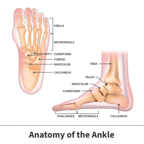
The ankle is a large joint connecting the leg and foot. Ankle joints also bring three key bones together. These are the shin bone (tibia), the leg bone (fibula), and the heel bone (talus).
The ankle and the area immediately surrounding it are made up of soft tissues like ligaments and tendons, muscles, blood vessels, and nerves.
About arthroscopy of the ankle
When the ankle or neighboring regions is injured or diseased, any number of conditions may develop that might eventually bring forth the need for ankle arthroscopy. Specific occurrences include:
- Impingement – This condition happens when soft tissues grow inflamed and press against or impinge on nearby structures.
- Arthrofibrosis – A stiffness or thickening of the ankle joint can result in arthrofibrosis. The problem often develops because scar tissue from some type of previous injury collects inside the ankle.
- Loose fragments – Occasionally, ankle injuries can lead to the loosening of small pieces of bone or soft tissues. When these small pieces travel, they can cause damage to things they encounter.
- Synovitis – The ankle joint is lined with a protective covering called synovial tissue. When this safeguard is injured or inflamed, problem symptoms can result. Often, the condition is caused by overuse of the ankle joint. It might also result from diseases like arthritis.
- Infection – Arthroscopy of the ankle may be used to treat infections. Soft tissue infections often require internal cleansing of diseased structures besides antibiotic treatment.
- Fractures – can fractures to ankle bones can be repaired with arthroscopy. Broken structures might be positioned back together or fused using this technique.
Symptoms
Symptoms of ankle issues that may need an arthroscopic procedure will range in appearance and severity, based on the specific problem. Many of these concerns produce common symptoms, including:
- Pain
- Discomfort that worsens when partaking in any movement or exercise
- Swelling
- Redness
- Bruising
Moderate or severe issues may cause instability (the feeling that the ankle joint is loose and might give out at any time), the inability to place weight on the wounded foot, and mobility limitations.
Possible complications
If not correctly addressed, certain ankle problems can eventually lead to chronic pain and serious mobility issues. Such issues might weaken other ankle, foot, or leg parts resulting in further injuries and associated symptoms.

Diagnosis
Your orthopedic specialist will often begin the diagnostic process by asking you several questions designed to give them a better idea of how to proceed. Such questions might include the following:
- When did the pain start?
- What type of work do you do?
- What leisure or athletic activities do you participate in?
- Do any movements make the pain worse?
- Were you previously diagnosed with an ankle, foot, or leg injury?
This first stage is usually followed by a complete physical evaluation where your physician will closely examine your ankle to identify any apparent abnormalities. They might also ask you to perform various movement tests to determine the pain threshold and motion ranges.
Internal ankle damage is often confirmed using diagnostic imaging tests such as X-rays, MRI (Magnetic Resonance Imaging) scans, or CT (Computerized Tomography) scans.
Arthroscopy of the ankle procedure
Ankle arthroscopy is a minimally invasive procedure used to treat specific ankle conditions.
Surgeons perform ankle arthroscopy using a small tube-like device called an arthroscope. It is equipped with a tiny camera capable of producing images that can be sent to a computer. Arthroscopes are also fitted with small but specially designed surgical tools.
Both doctors and patients favor this form of treatment because it requires smaller incisions, less bleeding, or any other potential concern associated with traditional open surgery and usually takes less time to recover.
Preparation
Patients might be asked not to eat or drink specific products in the days and hours leading up to the procedure. They are also advised to mention any over-the-counter or prescription medications they routinely take to their doctor. Certain drugs might interfere with the process or increase the possibility of experiencing post-surgical complications.
The process
Before beginning the procedure, the patient will be given some form of anesthesia. Once sedation has set in, the area surrounding the patient’s ankle will be cleaned and sterilized.
Surgeons then make small cuts to enable the arthroscope’s portals to enter. Once this step is complete, doctors will insert the device and perform whatever actions are necessary for the patient in question.
Once the procedure is complete, the arthroscope will be carefully removed. The last stage of the process involves surgeons stitching and bandaging the portal openings.
Recovery
Every patient’s recovery time differs and varies based on their age, weight, physical activity level, general health, the specific underlying condition for which the procedure was used to correct, and that issue’s severity. The healing process will likely need a period where the patient’s foot is immobilized to prevent re-injury and a potential course of physical therapy.
Videos
Related specialties
- Achilles Calcific Tendinitis
- Achilles Tendon Rupture
- Achilles Tendonitis
- Ankle Fracture Surgery
- Ankle Fractures (Broken Ankle)
- Ankle Fusion Surgery
- Arthroscopic Articular Cartilage Repair
- Bunions
- Charcot Joint
- Common Foot Fractures in Athletes
- Foot Stress Fractures
- Hallux Rigidus Surgery - Cheilectomy
- Hammer Toe
- High Ankle Sprain (Syndesmosis Ligament Injury)
- Intraarticular Calcaneal Fracture
- Lisfranc Injuries
- Mallet, Hammer & Claw Toes
- Metatarsalgia
- Neuromas (Foot)
- Plantar Fasciitis
- Sprained Ankle
- Total Ankle Replacement
- Turf Toe
