Heel bone fracture
Intraarticular calcaneal fracture
An intraarticular calcaneal fracture, or heel bone fracture, is a fracture that occurs in the heel usually due to a high-impact injury, such as a car accident or falling from a ladder. The severity of the fracture is dependent on the intensity of the impact during the time of injury; the more severe the injury, the more severe the fracture will be. In most cases, surgery is the primary treatment option, and if often followed by physical therapy and an intensive recovery process.
Anatomy

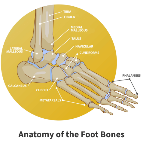
The bones of the feet are divided into three parts: the hindfoot, midfoot, and forefoot. Seven bones, known as tarsals, make up the hindfoot and midfoot. The calcaneus (heel bone) is the largest of the tarsal bones in the foot. It lies at the back of the foot (hindfoot) below the three bones that make up the ankle joint. These three bones are the:
- Shinbone (tibia)
- Smaller bone in the lower leg (fibula)
- Small foot bone that works as a hinge between the tibia and the fibula (talus)
Together, the calcaneus, heel bone, and the talus form the subtalar joint. The subtalar joint allows side-to-side movement of the hindfoot and is crucial for balance on uneven surfaces.
About heel bone fractures
An intraarticular fracture is a bone fracture in which the break crosses into the surface of a joint. This always results in damage to the cartilage. A calcaneal fracture, also known as a heel bone fracture, occurs when the heel is crushed under the weight of the body, like in a car crash or a fall from a ladder. The severity of an intraarticular calcaneal fracture can vary greatly. For example, a twisted ankle may result in a single crack in the bone, while something more high impact like falling from a ladder may cause the entire bone to shatter. The severity of a calcaneus injury depends on the following factors:
- The number of fractures
- The damage to the cartilage surfaces in the subtalar joint
- The amount and size of the broken bone fragments
- The damage to surrounding soft tissues, such as muscle, tendons, and skin
- The amount each piece is out of place (displaced) — In some cases, the broken ends of bones line up almost correctly; in more severe fractures, there may be a large gap between the broken pieces or the fragments may overlap each other.
When the bone breaks and the bone fragments stick out through the skin, the fracture is called an “open” fracture. An open fracture usually causes more damage to the surrounding muscles, tendons, and ligaments and takes longer to heal. Open fractures have a higher risk of infection in both the wound and the bone, so it is important for you to get immediate treatment to clean the wound and prevent an infection.
Symptoms
The most common symptoms associated with intraarticular calcaneal fractures include:
- Pain
- Bruising
- Swelling
- Limping
- Heel deformity
- Instability, causing you to walk differently
- Inability to put weight on the heel
- Inability to walk
Diagnosis
Your Florida Orthopaedic Institute physician will take a look at your symptoms and your overall health. It is important to tell your doctor if you have any other injuries or medical problems, such as diabetes, take medications or smoke, as they could be causing the problem. After discussing your symptoms and health, your physician will:
- Examine your foot and ankle to check if you have an open fracture.
- Check your pulse to make sure that you have good blood supply to the foot and toes.
- Check to see if you can move your toes.
- Check to see if you still have feeling on the bottom of your foot.
- Determine whether you have injured any other areas of your body
Your physician may also order some tests to ensure that you actually have an intraarticular calcaneal fracture and not another condition. These tests include:
- Computed tomography (CT) scans – This test produces a more detailed, cross-sectional image of your foot and provides your doctor with valuable information about the severity of your fracture.
- X-rays – This test creates images of dense structures, such as bone. It can show if your calcaneus is broken and whether the bones are displaced.

Treatment
Nonsurgical treatments are only recommended if the bones fractured remain in place. If they have moved out of place, then surgery will be necessary to put the bone back together. The healing process can take anywhere from 6 weeks to over 3 months, and is dependent on the severity of the fracture.
Nonsurgical treatment
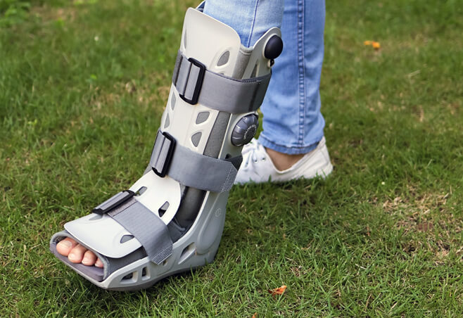
Nonsurgical treatments may be recommended if the pieces of broken bone have not moved out of place from the force of the injury. Immobilization is the only non-surgical treatment available. It involves using a cast, splint, or brace to hold the bones in your foot in the proper position for approximately 6 to 8 weeks. No weight can be put on your foot during this time to ensure a successful healing process.
Surgical treatment
There are several different surgical options available for intraarticular calcaneal fractures, including:
- Open reduction and internal fixation – During this procedure, an open incision is made to reposition the bones into their normal alignment. They are held together with wires or metal plates and screws.
- Percutaneous screw fixation – If the bone pieces are large, they can sometimes be moved back into place without making a large incision. Special screws are then put into the heel through small incisions to hold the fracture together.
- After your procedure, pain medications are often prescribed to handle the pain. Many types of medications are available to help manage pain, including opioids, non-steroidal anti-inflammatory drugs (NSAIDs), such as ibuprofen (Advil, Motrin) and aspirin, and local anesthetics. Your doctor may use a combination of these medications to improve pain relief, as well as minimize the need for opioids.
Recovery
Whether your treatment is surgical or nonsurgical, your rehabilitation process will be very similar. The time it takes to return to daily activities will vary depending on the type and severity of the fracture, and whether you have other injuries.
Some patients can begin weight-bearing activities a few weeks after injury or surgery. Others may need to wait approximately 3 months before putting weight on the heel. Most patients are able to begin partial weight bearing between 6 and 10 weeks after injury or surgery.
Many doctors encourage motion of the foot and ankle early in the recovery period. For example, you may be instructed to begin moving the affected area as soon as your pain allows. If you have had surgery, you may be instructed to start moving the affected area as soon as the wound heals to your doctor’s satisfaction.
Physical therapy may also be recommended to speed the healing process. Specific exercises can help strengthen supporting muscles and improve the range of motion in your foot and ankle. Although they are often painful at the beginning and progress may be difficult, exercises are needed in order for you to resume normal activities.
When you begin walking again, you may need to use a cane, walker, crutches, or even a special boot. If you put weight on your foot too soon, the bone pieces may move out of place and you might need surgery. If you have had surgery, the screws might loosen or break, and the bone may collapse. This may not occur the first time you walk on it but, if the bone is not healed and you continue to bear weight, the metal could eventually break, resulting in another surgery.
Videos
Related specialties
- Achilles Calcific Tendinitis
- Achilles Tendon Rupture
- Achilles Tendonitis
- Ankle Fracture Surgery
- Ankle Fractures (Broken Ankle)
- Ankle Fusion Surgery
- Arthroscopic Articular Cartilage Repair
- Arthroscopy of the Ankle
- Bunions
- Charcot Joint
- Common Foot Fractures in Athletes
- Foot Stress Fractures
- Hallux Rigidus Surgery - Cheilectomy
- Hammer Toe
- High Ankle Sprain (Syndesmosis Ligament Injury)
- Lisfranc Injuries
- Mallet, Hammer & Claw Toes
- Metatarsalgia
- Neuromas (Foot)
- Plantar Fasciitis
- Sprained Ankle
- Total Ankle Replacement
- Turf Toe
