LCL injuries
Lateral collateral ligament injuries
A lateral collateral ligament, or LCL injury happens when the ligament located in the knee joint is injured. Ligaments are thick, strong bands of tissue that connect bone to bone, which helps to keep the knee joint stable. A lateral collateral ligament injury does not have to be completely torn. It can also be strained, sprained, or partially torn. There are both surgical and nonsurgical treatments available to help heal your ligament injury. Nonsurgical treatments are the main way to treat the injury. But if the injury is severe, then surgery may be necessary.
Anatomy

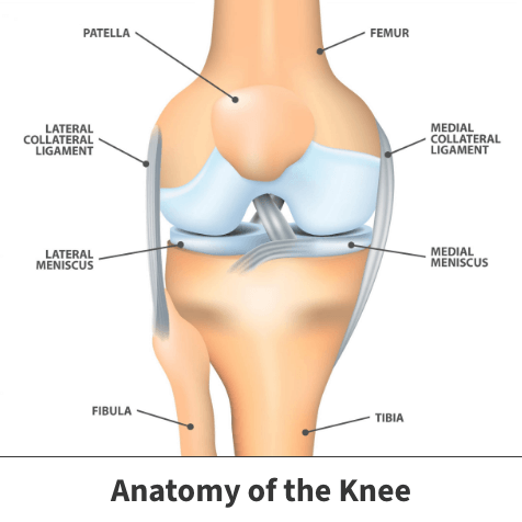
Three bones meet to form your knee joint: your thighbone (femur), shinbone (tibia), and kneecap (patella). Your kneecap sits in front of the joint to provide protection.
Bones are connected with ligaments. There are four primary ligaments in your knee. They act like strong ropes to hold the bones together and keep your knee stable. The lateral collateral ligament (LCL) runs along the outside of the knee joint, from the outside of the bottom of the femur to the top of the fibula. The LCL helps keep the knee joint stable, especially the outside of the joint.
About
A lateral collateral ligament injury is when the ligament located in the knee joint is injured. Since the knee relies solely on the surrounding ligaments and muscles for stability, it can be easily injured. Any direct contact to the knee or hard muscle contraction, such as changing direction rapidly while running, can injure a knee ligament.
An injury to a lateral collateral ligament may include straining, spraining, and partially or completely tearing any part of the ligament. The leading cause of lateral collateral ligament injuries is from a direct-force trauma to the inside of the knee, causing the knee to be pushed outward. This puts pressure on the outside of the knee and causes the ligament to stretch or tear.
Symptoms
There are several symptoms associated with lateral collateral ligament injury, including:
- Pain on the outside of the knee
- Swelling over and around the area of the injury
- Knee instability and weakness
Diagnosis
Your Florida Orthopaedic Institute physician will take a look at your symptoms and overall medical history, followed by a physical examination. During the exam, your physician will look at your injured knee and compare it with your non-injured knee. Most ligament injuries are diagnosed through a physical exam. Sometimes imaging tests may be needed to confirm the diagnosis. These tests include:
- Magnetic resonance imaging (MRI) scans –This test creates images of soft tissues like the collateral ligaments. If your lateral collateral ligament is torn, this test will show it.
- X-rays – While x-rays will not show any injury to your collateral ligaments, they can identify if you also have a broken bone.
Treatment
There are both surgical and nonsurgical treatments available to help get you back on your feet. Nonsurgical treatments are the main way to treat lateral collateral ligament injuries. But if the injury is unable to heal, then your physician may recommend surgical procedures.
Nonsurgical treatments
Icing, bracing, and physical therapy are the three main nonsurgical options that your Florida Orthopaedic Institute physician can recommend. Typically, non-surgical treatments are recommended first unless your symptoms are severe and need immediate surgical procedures.
- Ice – Icing your injury is crucial to the healing process. The proper way to ice an injury is to use crushed ice directly to the injured area for 15 to 20 minutes at a time, with at least 1 hour between icing sessions.
- Bracing – It is important for your knee to be protected from the same sideways force that caused the injury. Bracing helps protect the injured ligament from stress. To further protect the knee, you may also be given crutches which helps from putting weight on your leg. You may need to change your daily activities to avoid movements that could further injure your knee.
- Physical therapy – Physical therapy can restore function to your knee and strengthen the leg muscles that support it.
Learn More About Physical Therapy
Surgical treatments
Most lateral collateral ligament injuries can be treated successfully without any surgical treatments. But if the torn ligament is unable to heal or is associated with other ligament injuries, your Florida Orthopaedic Institute physician may suggest a surgical procedure to fix it.

Videos
Related specialties
- ACL Injuries
- Arthroscopic Chondroplasty
- Articular Cartilage Restoration
- Deep Thigh Bruising
- Fractures of the Tibial Spine
- Iliotibial Band Syndrome
- MACI
- Medial Collateral Ligament Injuries
- Meniscus Tears
- Muscle Spasms
- Muscle Strains of the Calf
- Partial Knee Replacement
- Patellar Fracture
- Quadriceps Tendon Tear
- Runner's Knee
- Senior Strong
- Shin Splints
- Total Knee Replacement Surgery
