Partial knee replacement
Knee replacement unicompartmental
Knee pain that doesn’t go away and keeps getting worse makes life harder. And, as more time passes without relief, frustration often increases—along with the discomfort—if nonsurgical treatments (like rest, physical therapy, medications, or injections) have failed to provide relief. For patients in situations like this, an orthopedic physician specializing in knee care might recommend a total or partial knee replacement to alleviate pain and improve mobility.
Keep reading for our partial knee replacement FAQ and learn more about:
- The anatomy of a knee,
- What a partial knee replacement is,
- Who benefits from this surgical knee repair and
- What to consider when choosing a knee surgeon.

Anatomy

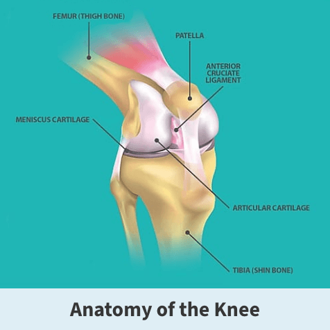
The knee is a hinge joint, meaning it can only bend in one direction, and is where the tibia, the femur, and the patella meet. Like most joints in the body, the knee has a dense, fibrous, connective tissue, known as articular cartilage, that seals the joint space between the femur and tibia, and prevents the bones from rubbing against each other. The articular cartilage acts as a shock absorber and allows for smooth and stable movement. It is divided into three major compartments:
- The inside part of the knee (Medial compartment),
- The outside part of the knee (Lateral compartment) and
- The front of the knee between the kneecap and thighbone (Patellofemoral compartment)
What is partial knee replacement?
Your knee has three compartments: the medial (or inside compartment), lateral (outside compartment), and patellofemoral (front compartment).
A partial knee replacement, or unicompartmental knee arthroplasty (UKA), involves fixing only one of the three compartments (leaving the other healthy compartments and ligaments as is). In contrast, a surgeon replaces all three compartments during a total knee replacement. To perform a partial knee, the anterior (ACL) and posterior (PCL) ligaments must be intact and in good condition.
This option often leads to a quicker recovery, provides a more natural feeling to the knee, maintains more bone and ligaments, and preserves more of your natural mobility. A patient is more likely to be able to return to sports if a partial knee replacement is performed.
How is the need for a partial knee replacement determined?
Part of the diagnosis process may involve taking X-rays to view cartilage loss and joint space narrowing. Your physician may also order MRI scans (if X-rays don’t show a definite reason for your joint pain or if your X-rays indicate that other joint tissue might be damaged) and blood tests (to rule out other potential conditions as the reason for your knee pain).
Your Florida Orthopaedic Institute physician will evaluate your symptoms, imaging results and circumstances to determine whether a partial knee arthroplasty is reasonable.
How long will my partially-replaced knee last?
A partial knee replacement’s lifespan depends on several factors, and implants will generally last for around:
- 10 years for 95% of patients,
- 15 years for 90% of patients, and
- 20 years for 80%: of patients.
Watch Dr. Brian T. Palumbo describe patient selection’s impact on partial knee replacement outcomes
What happens, step-by-step, during surgery to partially replace a knee?
At a high level, a total knee replacement surgery follows these four steps:
- During a partial knee replacement surgery, you’ll first receive anesthesia—either general (putting you to sleep) or regional (numbing the lower half of your body)—to ensure you’re comfortable and pain-free throughout the procedure.
- The surgeon then makes an incision in the front of your knee. They carefully cut and remove the diseased cartilage and bone from the single, particular arthritic compartment. (In the case of a total knee replacement, the surgeon replaces all three knee joint compartments.)
- Then, the surgeon caps the cut bone surfaces with a metal implant, effectively restoring the knee joint surface’s shape and dimension. A partial replacement offers a targeted approach that maintains more of your natural knee, often leading to a quicker recovery and providing a more natural feeling knee.
- Finally, once everything is securely in place and aligned correctly, the surgeon will close the incision and apply a dressing to protect the wound.
Immediately after surgery, you’ll start putting weight on your new knee, usually with the help of a walker, cane, or crutches. A physical therapist will give you exercises to help maintain your range of motion and restore your strength during healing.
See how a personal trainer is back to helping others after two knee replacements

What’s life like after a partial knee replacement?
Partial knee replacements require approximately six weeks of recovery, which includes specific rehabilitation exercises to facilitate healing. Fortunately, because this procedure is less invasive, people usually recover faster than after total knee replacement surgery.
Following your doctor’s care instructions also contributes to a smooth healing process. The recovery phase of a partial knee replacement typically involves four to six weeks of physical therapy to help restore movement and strengthen the knee.
Most patients begin walking with assistance soon after the surgery and gradually increase their activity levels. While recovery times can vary, many individuals can resume their daily activities within two to four months, experiencing improved mobility and reduced pain. Following prescribed exercises and attending regular follow-up appointments with your healthcare team are essential for a successful recovery.
Hear Dr. Spencer Smith talk about how he helps patients with failed knee replacements
What to expect after partial knee replacement surgery
Beyond one year, partial knee replacement was considered more cost-effective than total knee replacement, according to research published in the National Library of Medicine. A partial replacement was also associated with more significant health benefits (measured using quality-adjusted life years) and lower healthcare costs, reflecting lower costs of the index surgery and subsequent healthcare use.
Potential complications and risks associated with partial knee replacement
One potential downside to this procedure is the potential for future surgery and conversion to a total knee replacement. For example, if the knee you had a partial replacement on deteriorates in the future, you may need to have a total knee replacement. This is a somewhat complex decision, and you should discuss it with your surgeon.
Partial knee replacement operations are more challenging than total knee replacements and are more susceptible to implantation errors. Patients who are good candidates for successful partial knee replacements weigh less than 220 pounds, have healthy ACLs, and their other knee compartments are healthy.
Watch Dr. Brian T. Palumbo describe patient selection’s impact on partial knee replacement outcomes
Who benefits from partial knee arthroplasty?
The knee is a hinge joint, meaning it can only bend in one direction. It is where the tibia, the femur, and the patella meet. Like most of the body’s joints, the knee is lined with articular cartilage. The articular cartilage acts as a shock absorber, allowing smooth and stable movement. When the articular cartilage wears away, knee replacement can alleviate pain and restore movement and function.

Signs you might need a partial knee replacement
Some of the general symptoms associated with a need for a partial knee arthroplasty include:
- Pain that increases with activity but only gets slightly better with rest,
- A feeling of warmth in your knee joint,
- Stiffness in the knee, especially in the morning or after sitting for long periods,
- Decrease in knee mobility (that makes it difficult to get in and out of chairs or cars, use the stairs or walk),
- Swelling, and
- Creaking or crackly sound when your knee moves.
If you’re struggling with severe knee pain and limited movement from arthritis or joint damage from other causes, knee arthroplasty may be the right treatment to help improve your quality of life.
Developments in surgical practices, coupled with innovative technologies, make the replacement of your entire knee one of the most successful surgeries. In fact, total knee replacement surgeries continue to grow yearly, with almost a million successful replacements each year in the United States alone.
Discover how knee replacement at FOI helped one biker ride his Harley once more
Advances in surgery for knee replacement
Since the first total knee replacement surgery was performed in 1968, surgery to replace the knee joint partially and completely has continually evolved in its usefulness, effectiveness, and overall success. Recent knee surgery innovations that have significantly enhanced patient outcomes and recovery experiences include:
- Augmented reality
- NAVIO
- MAKO
- Pain management advancements
One is computer navigation, which can guide the placement of several partial knee replacement implants. However, the medical literature still debates whether this computer guidance offers substantial benefits. That said, if a surgeon doesn’t perform many partial knee replacement procedures, using computer guidance may help with implant precision and alignment.
At Florida Orthopaedic Institute, our surgeons use several computer guidance systems, including augmented reality (ARVIS—Enovis) and surgical robots (NAVIO and MAKO).
Augmented reality
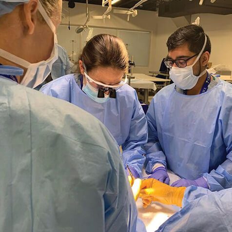
ARVIS is an augmented reality platform from Enovis. To operate it, a surgeon wears a helmet with special glasses that provide a heads-up display for communicating with a computer navigation system. The glasses visualize anatomy and input from the surgical field and provide real-time objective feedback to facilitate bone cuts, implant position, balance, and alignment.
A surgeon getting assistance from ARVIS during joint surgery is reminiscent of JARVIS supporting Tony Stark while wearing his Iron Man suit.
NAVIO
The NAVIO surgical system is a robotics-assisted navigation technology that consistently and accurately positions the knee implant. It does this by building a 3D model of the patient’s knee using anatomical data provided by a surgeon. This technology for knee replacement allows a surgeon to place the implant and balance the knee’s ligaments for optimal alignment and a well-balanced knee.
NAVIO allows surgeons to take advantage of the patient’s unique anatomy and tailor the procedure to better suit the patient’s needs. This offers many benefits to the patient, such as a smaller incision, less pain, quicker rehabilitation and recovery, more natural knee motion, and a lower risk of complications.
MAKO
A surgeon can create a detailed, pre-operative plan for your partial knee replacement by loading a 3D model—specific to your unique anatomy based on a CT scan of your knee—into the MAKO surgery system.
Hear Dr. Kenneth A. Gustke speak on other benefits of the MAKO surgery system

Pain management advancements
New pain management techniques, including improved anesthesia protocols and targeted pain control strategies, help minimize post-operative discomfort and enable patients to begin physical therapy sooner.
Combined, these innovations make knee replacement surgeries safer, more efficient, and more comfortable, allowing patients to regain mobility and quality of life more smoothly.
Hear Dr. Hari Parvataneni share why he considers patient relationships as partnerships
Selecting a unicompartmental knee arthroplasty surgeon
Finding a great knee physician is essential for a successful replacement. Take your time to research potential knee arthroplasty providers by evaluating the following crucial factors that will help you make an informed decision:
- Experience and expertise: Seek out a knee replacement surgeon with a meticulous knowledge of implanting components correctly during knee arthroplasty and a high patient satisfaction score. You’ll also want to choose a surgeon who performs over 30 partial knee replacements yearly. You might also want to pick someone with certification or training in using advanced technologies (such as augmented reality or robotics guidance systems).
- Convenient care options: Make your life easier. It’s often advantageous to pick a surgeon in a robust orthopedic health system (with various locations and comprehensive services) because this can make scheduling more flexible and coordinate related care needs (such as imaging, physical therapy, and rehabilitation).
- Patient feedback: For referrals, consult trusted friends, family members, or your primary care provider. Researching reviews and reading patient testimonials is also an excellent way to understand others’ experiences with a provider during knee replacement surgery.
Discover how knee replacement at FOI changed a U.S. Army vet’s life for the better
When to see a specialist about partial knee replacement
Deciding whether to undergo knee replacement is a significant step. Although it’s considered a safer orthopedic procedure, total knee replacement is still a major operation, so it’s natural to question whether it’s right for you.
Here are examples of when it might be time to think about having complete knee replacement surgery:
- You’ve exhausted conservative treatment options without satisfactory relief.
- Your pain and loss of knee function make it hard to perform everyday tasks and enjoy activities you love.
- You’re in good overall health and medically fit for surgery—able to tolerate anesthesia and rehabilitation.
- You’re mentally prepared for total knee replacement surgery and motivated to participate in the post-operative recovery process.
If you think having knee replacement surgery might be right for you, schedule an appointment to discuss your symptoms, medical history, and treatment options with an experienced knee surgeon.
Find out how a custom knee implant helped a 77-year-old CEO golf again
Why choose FOI for partial knee replacement
Unicompartmental knee replacement surgery is considered one of the most successful medical operations. Here at Florida Orthopaedic Institute, our fellowship-trained knee surgeons are renowned specialists who combine their world-class training with innovation to provide patients with individualized solutions designed to help them stay active.
Picking the right surgeon is essential for a successful knee replacement experience. Our team is here to support you with expertise, convenience, and comprehensive care. When you’re ready, contact us to schedule a consultation.
During this appointment, you can discuss partial knee replacement options with a surgeon and determine the best surgical approach for your needs.
Listen to an active retiree reveal what life is like after having two knee replacements

Partial knee replacement FAQs
Even though a partial knee replacement can significantly reduce pain and improve function, your knee may not feel exactly how it did before you started experiencing pain and losing mobility. That said, partial knee replacements are more likely to feel more natural than total knee replacements.
To help your partial knee replacement last longer, follow your knee surgeon’s post-op advice, keep a healthy weight, stay active with easy exercises, and attend regular check-ups.
Hear Dr. Steven T. Lyons discuss the features of Smart Knee medical implants
Yes. Generally, patients who undergo partial replacement experience less pain and quicker recovery since the surgery is less extensive and only part of the knee is replaced.
Surgeons typically make a smaller incision for a partial knee replacement, usually around three to five inches long. This cut is much smaller than the one made during total knee replacements (which may require an incision eight inches or longer) and helps reduce tissue damage, leading to a quicker recovery.
Listen to Dr. Grant G. Garlick talk about outpatient knee replacements at FOI
Watch Dr. Thomas L. Bernasek describe same-day joint surgery’s benefits
There is no specific “best” age for a partial knee replacement. It’s more about your overall health and activity level. Many active people in their 50s and 60s find them beneficial.
Common mistakes after a partial knee replacement include skipping physical therapy, not following your doctor’s instructions, and pushing yourself too hard too soon. Stick to your rehab plan and listen to your body to ensure a smooth recovery.
After partial knee replacement, most people go home the same day or after a short hospital stay and start walking with assistance right away. Full recovery typically takes a few weeks to a few months, depending on your health and commitment to physical therapy.
Red flags after a partial knee replacement include severe pain, excessive swelling, redness or warmth around the knee, fever, and any unusual discharge from the incision site. If you experience these symptoms, contact your doctor immediately.

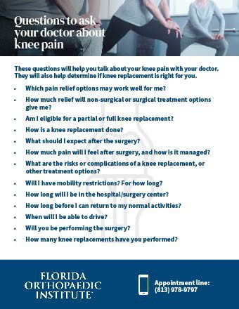
Should you see a specialist about your knee pain? Answer these questions to find out.
Videos
Related specialties
- ACL Injuries
- Arthroscopic Chondroplasty
- Articular Cartilage Restoration
- Deep Thigh Bruising
- Fractures of the Tibial Spine
- Iliotibial Band Syndrome
- Lateral Collateral Ligament (LCL) Injuries
- MACI
- Medial Collateral Ligament Injuries
- Meniscus Tears
- Muscle Spasms
- Muscle Strains of the Calf
- Patellar Fracture
- Quadriceps Tendon Tear
- Runner's Knee
- Senior Strong
- Shin Splints
- Total Knee Replacement Surgery
